


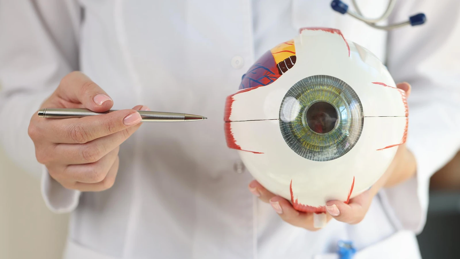
The structure of the eye has always been a fascinating topic to discuss. Beyond all the usual talk about eye colour, what truly makes them interesting is their intricate and detailed structure. The eye is a small yet very important part of the body; it helps us visualise the beauty of this world.
In this modern era, our eyes are under various kinds of threats from digital rays; hence, an acknowledgement of their structure and function helps to comprehend them efficiently. Do you ever wonder what light does to our eyes to let us see any object clearly? If not, we’re here to walk you through the full structure of the eye and its functions to help you understand it more clearly.
How Does the Structure of the Eye Work?
Though the structure of the eyeball looks like a sphere, it is not exactly a sphere. The structure of the eye consists of two units called the anterior segment and the posterior segment.
Cornea, lens and iris make up the anterior segment, while the cornea, being a transparent and curved part, also forms a part of the posterior segment. The posterior segment is the back portion or two-thirds portion of the eye that is composed of the anterior hyaloid membrane and optical structures like vitreous humour, optic nerve, retina and choroid. Iris is the pigmented circular part of the eye in the centre.
The black coloured looking structure is a pupil that predominantly controls the number of light rays entering our eyes with the help of the iris dilator and sphincter muscles. The light rays enter the eye through the cornea and through the pupil, and then through the lens. The lens changes shape for accommodation, which is controlled by the ciliary muscles.
A Guide to the Structure of Eyes
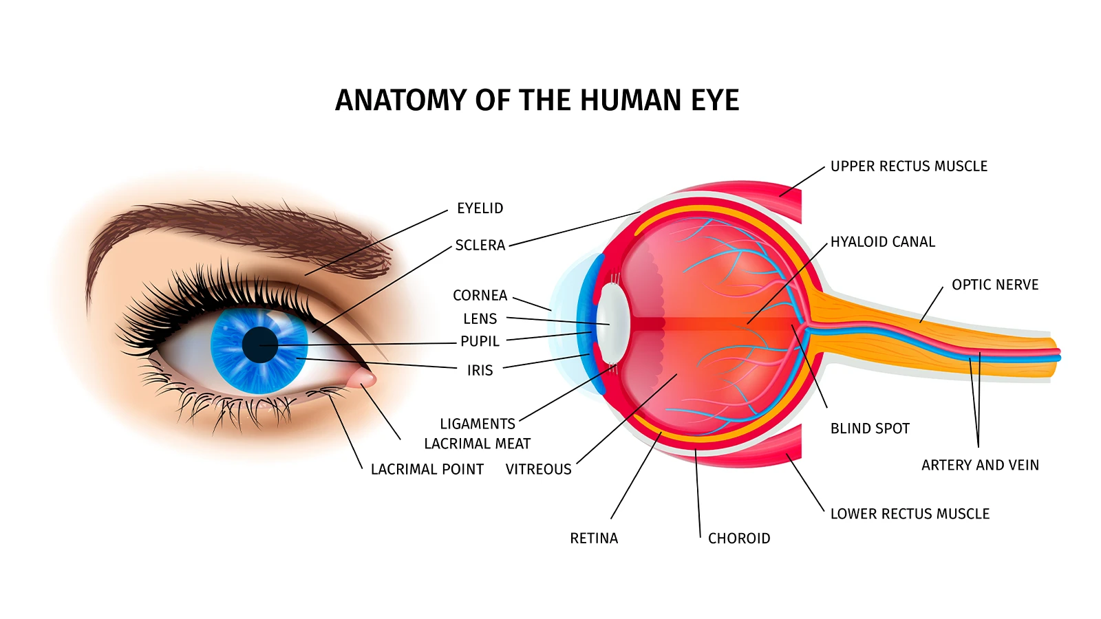
Vitreous Body - The vitreous body is a clear gel or glass-like gel that fills the gap between the lens and retina. The function of the vitreous is mainly to protect the eye. And most importantly, the fluid supports and holds the eyeball's spherical shape.
Cornea - It is a transparent part of the eye present at the very front that covers other parts like the iris, pupil, and anterior chamber. It regulates and focuses the entry of the light rays. The cornea decides the excess and lesser amount of light by either bending or reflecting the incoming light into the lens.
Iris - It is a thin and circular structure composed of two different layers called fibrovascular layers, also known as stroma and a pigmented epithelial cell beneath the other. Regulating the aperture of the pupil, the iris helps in the number of light rays entering the retina. Our eye colour is also defined by the iris.
Pupil - It is a hole in black colour located in the centre of the eye. It is responsible for regulating the amount of light in the eyes and then focusing it on the retina.
Ciliary Muscle - It is a ring of smooth muscles that is present above the lens and produces aqueous humour. The ciliary muscle controls the accommodation of objects at different distances.
Lens - The crystalline structure of the eye lens is a transparent structure present behind the iris that, along with the retina, helps to focus light on the retina.
Anterior Chamber - It is an aqueous humour-filled space inside the eye lying between the iris and the cornea's innermost surface, called the endothelium.
Conjunctiva - It is a tissue that lines the inner side of the structure of the eyelids and covers the sclera. The main function of the conjunctiva is to keep the front surface of the eye moist and lubricated, as well as the inner surface of the eyelids.
Sclera - It is a tough and dense white cover over the structure of the eyeball, along with the cornea, which forms the external coat. It is also known as the white of the eye.
Retina - It is a light-sensitive tissue at the back of our eye. The cornea, pupil and lens focus the light rays on the retina to form an image.
Macula - It is a part of the retina at the back of the eye. It is made mainly of cone cells that are responsible for the colour, central and sharp vision.
Optic Nerve - The optic nerve is the second of various cranial nerves that are responsible for bringing information from the retina to the brain.
Wrapping It Up
The human eye is a finely tuned organ where each part plays a vital role in vision. Understanding its structure and function helps us appreciate how we see and why eye care matters. Regular checkups and proper protection can preserve this remarkable sense for a lifetime. Wearing trendy eyeglasses can protect your eyes from dust, UV rays and external elements while adding a stylish touch. Good eye care blends health, comfort, and fashion effortlessly.
Caution: You may become style obsessed

Your way finder

2000+ Trendy Styles

Fashion Forward Sunnies


